XUV to X-ray radiation has characteristics that make it particularly attractive for imaging and in particular 3D imaging. On the one hand, the short wavelength (0.01 nm to 50 nm typically) makes it possible to improve the spatial resolution compared to visible imaging by greatly decreasing the diffraction limit. On the other hand, for wavelengths typically less than 1nm, matter begins to become transparent, regardless of its nature. It should be noted that there is a reduced spectral range, between 2.2 and 4.4 nm, for which water is transparent while carbon is still absorbent. This spectral window known as “water” makes it possible to image biological cells with high contrast. The transparency of matter paves the way for non-destructive 3D imaging. Finally, in a manner equivalent to the decrease in the diffraction limit, XUV and X-ray radiation makes it possible to achieve shorter pulse durations than in the visible range, typically attosecond (1 as = 10-18 s) and potentially zeptosecond (1 zs = 10-21 s). With this in mind, for several years now, we have been studying new 3D and ultra-fast XUV and X-ray imager schemes.
XUV Holography and XUV Diffraction: High-order harmonics as well as injected XUV lasers are sources with very high degrees of spatial coherence. High-order harmonics have also demonstrated the possibility of producing attosecond pulses.
In 2006, we performed one of the first holography demonstrations performed with a high-order harmonic bundle [A. S. Morlens et al., Optics Letters, 31, 21, pp. 3095-3097 (2006)]
In 2013, we studied the impact of wavefront defects of the high-order harmonic source on the quality of an XUV diffraction image. [X. Ge et al, Optics Express, 21, 9, pp.11441-11447 (2013)]
In 2015, we performed the first holography experiment with a beam containing a train of attosecond pulses. [G. Williams et al., Optics Letters 40, 13, 3205-3208 (2015)]
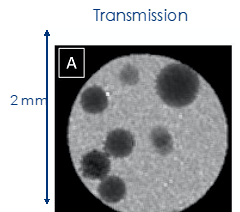
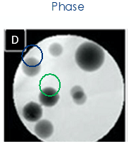
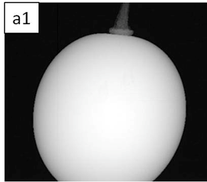
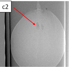
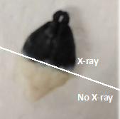
X-ray tomography: The reference technique in 3D X-ray imaging is still X-ray tomography, more commonly known as a scanner. Conventional algorithms for processing raw tomography data do not allow the separation of the real part from the imaginary part of the refractive index. However, each of these parts contains specific information about the material being probed. It is therefore interesting to be able to measure them separately
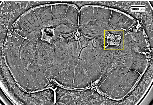
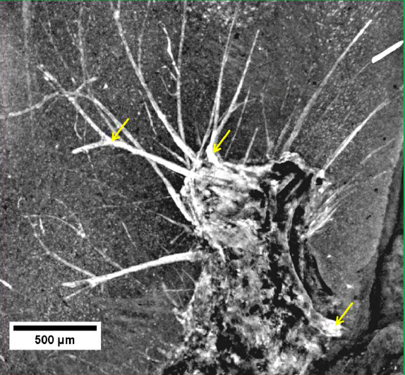
In 2018, we performed X-ray tomography experiments to estimate the distribution of radiosensitizing nanoparticles in healthy and tumor tissues of mice. (fig.1 and 2) [E. Longo et al., Journal of Instrumentation, 13 (2018)] [X. Le Guevel, Nanoscale, 39, 18657-18664 (2018)]