
Radiation biology with laser-accelerated particles
Laser-created plasmas can be used as a medium for particle acceleration. Thanks to the extremely high accelerating gradients that are formed in the high intensity laser-matter interaction, short and bright bunches of ionizing radiation (electrons, proton, X-ray photons) are produced. These radiation qualities maintain the signature of the acceleration phenomenon at their origin: the initial repetition rate and the very short duration.
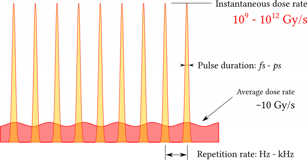
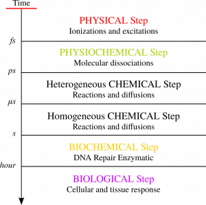
Figure 1: Left: different phenomena involved in ionizing radiation interaction with living organisms. Right: temporal structure of dose deposition by laser-accelerated particles
The biological effect of ionizing radiation depends primarily on the absorbed dose, which is the amount of deposited energy (Gray : joules/kg). Recent studies have pointed out that not only the total deposited dose but also the temporal profile of its deposition do have a role in defining the whole biological effect. This evidence could be related to the number of disparate phenomena that are involved in the toxicity of ionizing radiation, taking place at disparate temporal (Fig.1-left) and spatial scales.
At the LOA we use laser-accelerated particles to study the effect of ultra-fast dose deposition at ultra-high dose-rates. This direct application of laser-accelerated particles enable us to explore the biological effect of ionizing radiation in the peculiar conditions produced by laser-driven sources: dose deposited as a sequence of ultra-intense and short radiation flashes (Fig.1-right).
Ad-hoc strategies are to be designed and implemented to transport and shape the accelerated beam, to precisely caracterize its uniformity and its stability and to mesure the deposited dose in a reliable way. This point in particular represents a non-negligible effort because the very nature of the produced beams (short duration, high intensity) demands adapted and non-conventional measurement protocols.
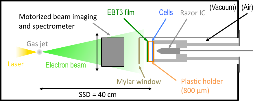
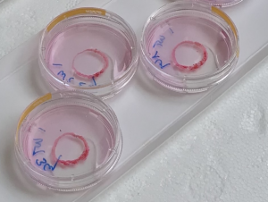
Figure 2: Left: setup for in-vitro experiment with low energy electrons. Right: Capsules with cell coltures
During the past few years both protons and electrons have been successfully used to irradiate cell monolayers. Survival assays (Fig.3-right) are used as a initial confirmation of the implemented dosimetry protocol. The surviving fraction of cells which received an increasing amount of dose usually follows a well known curve and remains stable from a campaign to the following one.
By varying the delay between consecutive shots (Fig.3-left) at constant deposited dose we have observed a variation of the surviving fraction. This effect is a consequence of a complex interplay between the timing of the radiation fraction and that of cell reparation mechanisms which resulted in more or less toxic irradiation conditions. Moreover two different mutations of the same cell line proved to behave in very different ways upon this variation. Such evidence motivates the quest for irradiation protocols able to increase the differential response between healthy and cancerous cells, towards a better comprehension of the toxicity mechanisms and the definition of novel cancer treatment strategies.
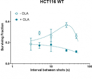
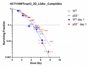
Figure 3: Left: Varying surviving fraction of irradiated cells with 7 MeV protons at constant dose and variable delay between laser driving pulses. Right: cell survival fraction after irradiation with 5 MeV electrons
Related publications:
L.Pommarel et al., “Spectral and spatial shaping of a laser-produced ion beam for radiation-biology experiments,” Phys. Rev. Accel. Beams, vol. 20, no. 3, p. 032801, Mar. 2017, doi: 10.1103/PhysRevAccelBeams.20.032801.
E.Bayart et al., “Fast dose fractionation using ultra-short laser accelerated proton pulses can increase cancer cell mortality, which relies on functional PARP1 protein,” Scientific Reports, vol. 9, no. 1, Dec. 2019, doi: 10.1038/s41598-019-46512-1.
M.Cavallone, A. Flacco, and V. Malka, “Shaping of a laser-accelerated proton beam for radiobiology applications via genetic algorithm,” Physica Medica, vol. 67, pp. 123–131, Nov. 2019, doi: 10.1016/j.ejmp.2019.10.027.
M.Cavallone et al., “Dosimetric characterisation and application to radiation biology of a kHz laser-driven electron beam,” Appl. Phys. B, vol. 127, no. 4, p. 57, Apr. 2021, doi: 10.1007/s00340-021-07610-z.
Contact: Alessandro Flacco
People involved: Alessandro Flacco, Jérôme Faure, Aline Vernier, Lucas Rovige, Joséphine Monzac